To find out about the most suitable vision correction option, register for a visit, or find out other important information, leave your contacts, and we will contact you as soon as possible.
Corneal transplantation
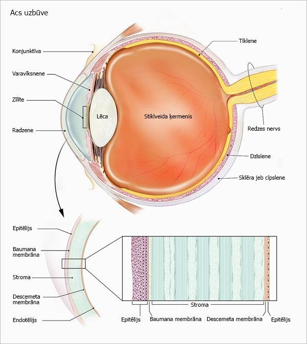
The cornea is the anterior, transparent part of the outer fibrous sheath of the eye. The cornea is one of the eye's optical system's main components - it focuses light rays to form an image inside the eye - on the retina.
The cornea looks transparent, but it has a complex, highly organized tissue structure.
There are no blood vessels in the cornea.
The cornea consists of 3 base layers, between which there are two thinner barrier layers or membranes:
The epithelium is the top layer, the outer layer, which protects the eye from foreign bodies - dust, water, bacteria. The epithelium absorbs oxygen and nutrients from the tears, thus providing nourishment to the cornea's deeper layers. There are thousands of fine nerve fibres in the epithelium, so the eye hurts if the cornea is crushed or scratched or if a foreign body enters the eye;
Bowman's membrane - behind the epithelium's basal cells is a thin layer of transparent tissue - Bowman's membrane. It is made of collagen fibres. If this membrane is damaged, it will heal and scar, which can lead to vision loss;
The stroma - the thickest - base layer of the cornea, consists mainly of collagen and water. Collagen gives the cornea strength, elasticity and shape. The unique structure of the corneal stroma ensures image quality and light focus in the eye;
Descemet's membrane - behind the stroma is a very thin but durable layer of tissue - Descemeta membrane, which acts as a protective barrier against infections and injuries. After damage, the Descemet's membrane may regenerate;
Endothelium - the inner layer of the cornea. It is a thin layer of cells that nourishes corneal tissue and keeps the cornea clear. Its primary function is to absorb excess fluid from the cornea. If the endothelial cells do not function well enough, the cornea becomes thick and no longer clear. There is a balance between the amount of fluid that enters the cornea and the fluid that is "pumped out" in the whole eye.
Unlike Descemet's membrane, endothelial cells damaged by trauma or disease do not regenerate.

Corneal transplantation
If the cornea is damaged and has a defect that cannot be cured with the medicine, there is a possibility that corneal transplantation surgery (keratoplasty) is needed.
Corneal transplantation or keratoplasty is the replacement of damaged corneal tissue with healthy donor tissue.
Transplantation can also be done for cosmetic purposes to remove a severe focal infection or maintain the eyeball's integrity at the corneal perforation.
Types of corneal transplants
There are two corneal transplantation types - complete corneal transplantation or penetrating keratoplasty (PKP) - the entire cornea (all layers) is replaced with donor tissue.
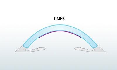
The second type is layered or lamellar corneal keratoplasty - only the damaged corneal layer is replaced with donor tissue. Descemet's Membrane Endothelial Keratoplasty (DMEK) is a deep lamellar keratoplasty - transplantation of the cornea's inner layers (Descemet's membrane and endothelium).

Descemet’s Membrane Endothelial Keratoplasty (DMEK) is one of the newest and most revolutionary corneal transplantation methods. The patient's corneal endothelium is removed and replaced with a donor endothelium. The donor membrane is attached to the cornea by an air bubble inside the eye. The tissue "clumps" quickly, but the air is absorbed into the tissue.
Advantages of DMEK operation:
- after DMEK surgery, patients can regain 90-100% visual acuity if there are no accompanying, impairing diseases;
- healing and rehabilitation time is relatively short - a few weeks;
- there are no sutures; thus, no high degree of astigmatism (corneal surface irregularity) is created;
- relatively low risk of rejection (corneal stromal tissue is not transplanted);
- the integrity and mechanical strength of the eyeball are maintained.
Compared to penetrating keratoplasty, DMEK is a much more gentle and less invasive method.
It has significantly improved transplantation outcomes in patients with corneal endothelial damage.
The clinic provides the necessary transplants for corneal transplant operations in cooperation with a living tissue bank in the United States.


